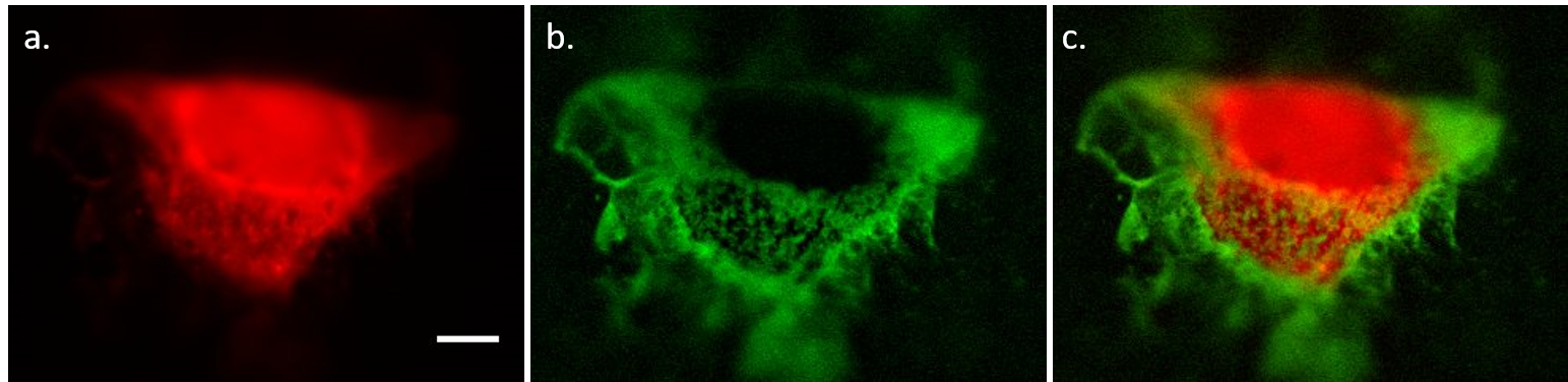
The development of novel single cell microscopy approaches continually opens avenues in bio-diagnostics. By labelling membrane proteins with fluorophores, photoluminescence (PL) has become the standard of single cell microscopy techniques. Despite the high sensitivity of PL methods, those techniques still present some inconveniences (e.g. high background due to autofluorescence). In recent years, the employment of chemiluminescence and bioluminescence methods has overcome PL challenges. In particular, electrogenerated chemiluminescence (ECL), thanks to the dual nature of electrochemical and photophysical phenomenon, offers many advantages: a higher selectivity, a very low background, and spatial resolution. The coreactant-based ECL system [Ru(bpy)3]2+/Tripopylamine (TPA), operating in buffered aqueous solutions, has permitted the wide diffusion of ECL as a bioanalytical technique.1 A previous study, based on [Ru(bpy)3]2+-labeled microbeads with TPA as coreactant, permitted to clarify the mechanism involved in bead-based immunoassays when the ECL-active species is immobilized as label.2 The knowledge of the mechanistic route represents the keystone of single cell ECL imaging. Chinese Hamster Ovary (CHO) cells were grown on a glassy carbon electrode, fixed and incubated with biotin X, which is able to react with primary amino groups of proteins. Then, biotin was linked with a streptavidin – modified [Ru(bpy)3]2+ label. The ECL luminophore [Ru(bpy)3]2+ can thus react with the electrochemically oxidized TPA species in solution to generate the excited state [Ru(bpy)3]2+* that emits light. In Fig. 1a, PL image shows the distribution of the labelling system at the level of the whole cell. ECL image (Fig. 1b) is then recorded by applying an anodic potential in a TPA buffered solution. By contrast with PL, ECL emission is mostly localized at cell membrane borders (see PL and ECL overlap in Fig. 1c). Because of ECL mechanism, light emission can happen only a few micrometers away from electrode surface.3 Overall, these studies demonstrate the feasability and possibilites of single cell imaging by ECL.
| Subject : | : | Oral (20 min) |
| Topics | : | ECL1. Electrochemiluminescence: from fundamentals to analytical applications |
| Keywords | : | Electrochemiluminescence ; Microscopy ; Cells ; Imaging |
| PDF version | : |  PDF version PDF version |
- Presentation
- Picture